Out of Print
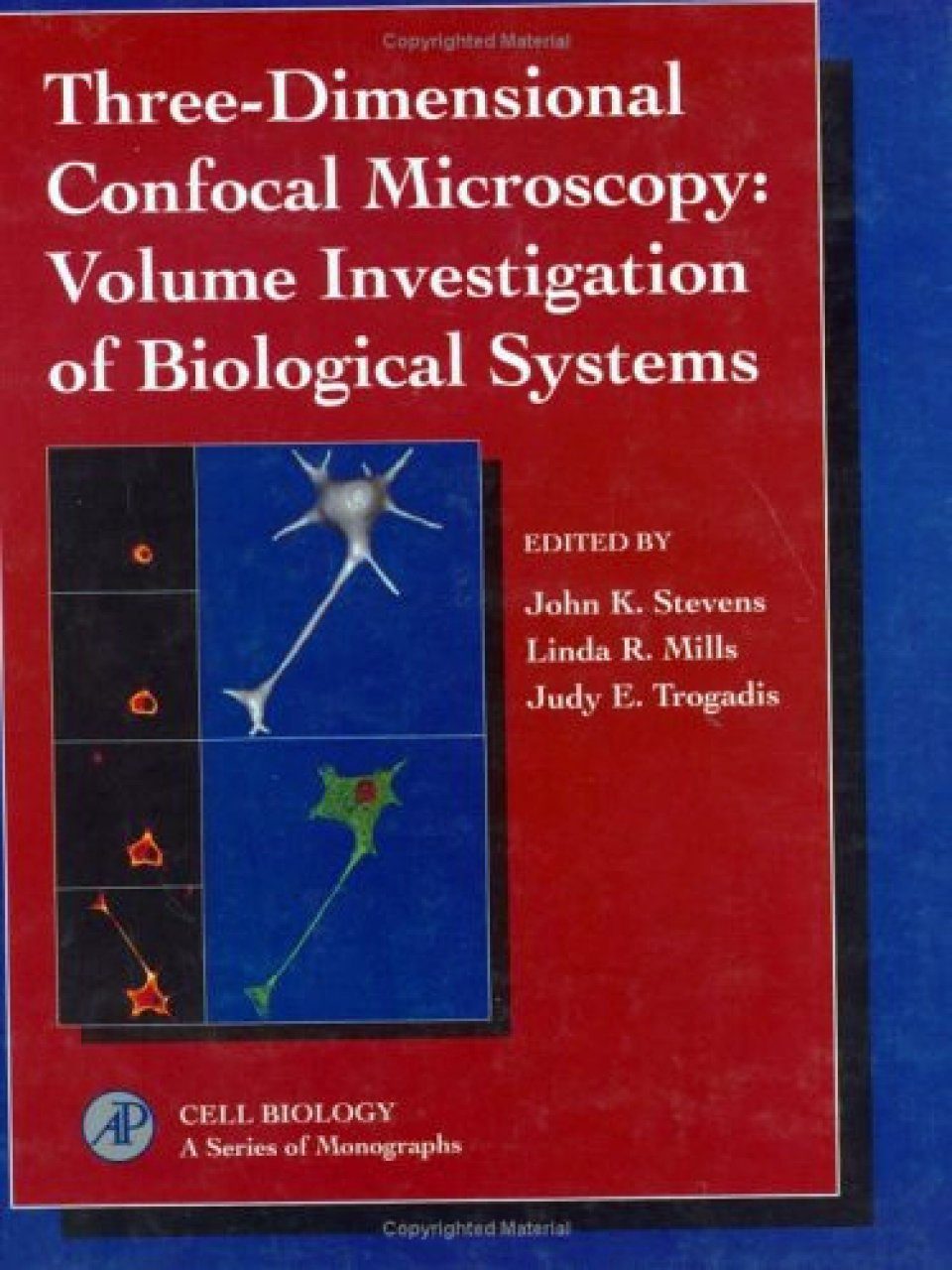
Edited By: JK Stevens, LR Mills and JE Trogadis
508 pages, Col & b/w photos, tabs, figs

Click to have a closer look
About this book
Contents
Biography
Related titles
About this book
The integration of confocal microscopy and volume investigation has led to an unprecedented ability to examine spatial relationships between cellular structure and function. This volume demonstrates their applicability to a wide range of biological and clinical problems.
Contents
Preface. Confocal Microscopy: Practical Considerations: Introduction to Confocal Three-Dimensional Volume Investigation. Background Rejection and Optimization of Signal to Noise in Confocal Microscopy. Sources of Noise in Three-Dimensional Microscopical Data Sets. Simultaneous Ultraviolet and Visible Wavelength Confocal Microscopy. Fluorescent Labels for Confocal Microscopy. Display Methods for Gray-Scale, Voxel-Based Data Sets. Three-Dimensional Volume Reconstitution in Confocal Microscopy: Practical Considerations. Biological Applications of Confocal Microscopy: Imagine Ion Channels in Live Central Neurons Using Fluorescent Ligands. Imaging Ion Channels in Live Central Neurons Using Fluorescent Ligands II. Labeling of Cells and Tissues. Expression of G Protein-Coupled Receptors n Baculovirus/Sf9 Cells: Imaging Receptor Distribution by Confocal Fluorescence Microscopy. Dynamic Intracellular Calcium Compartments: Confocal Microscopy Using Fluo-3 in Cells and Organelles. Confocal Microscopy of Living Eggs and Embryos. Spatial Organization of Microtubules in PC12 Cells: Three-Dimensional Electron Microscopy and Confocal Microscopy Applied to Volume Investigation. Confocal Imaging of Living Neurons and Organelles. Confocal Microscopy in Diagnostic Cytology. Confocal Microscopy in Diagnostic Pathology. Alternative Approaches and Future Applications: Ultrathin Optical Sectioning and Dynamic Volume Investigation with Conventional Light Microscopy. Serial Electron Microscopy as an Alternative or Complement to Confocal Microscopy for the Study of Synapses and Dendritic Spines in the Central Nervous System. Confocal Scanning Opthalmoscopy: Applying Laser Scanning Technology to Retinal Imaging. Use of Three-Dimensional Reconstructed Magnetic Resonance Imaging Data for Neurosurgical Planning. Index.
Customer Reviews
Biography
Ian Cameron is Reader in Chemical Engineering at the University of Queensland with teaching, research, and consultancy interests in process systems engineering. He has a particular interest in process modeling, dynamic simulation, and the application of advanced numerical methods to large-scale process systems. He has developed innovative courses in process modeling, design, and risk management, having extensive industrial experience in these areas. He continues to work closely with industry and government on systems approaches to process and risk management problems, as well as the development of internet-based tools for process systems applications.
Out of Print
Edited By: JK Stevens, LR Mills and JE Trogadis
508 pages, Col & b/w photos, tabs, figs


![Manuale di Microscopia dei Funghi [Manual to Microscopy of Fungi]](http://mediacdn.nhbs.com/jackets/jackets_resizer_medium/26/260752.jpg?height=150&width=104)

![Manuale di Microscopia dei Funghi, Volume 2 [Manual to Microscopy of Fungi, Volume 2]](http://mediacdn.nhbs.com/jackets/jackets_resizer_medium/23/236960.jpg?height=150&width=107)