United States
£ GBP
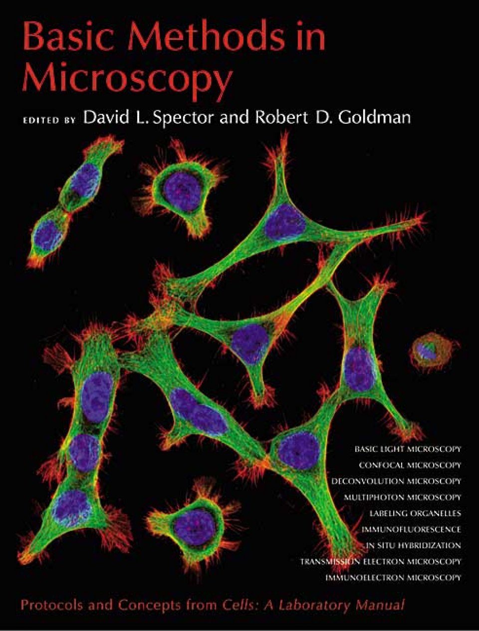
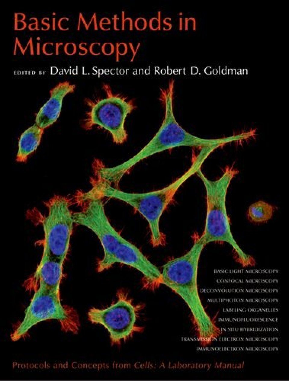
Imaging has become a vital tool for researchers in all aspects of biology. Recent advances in microscope technology, labeling techniques and gene and protein manipulation methods have led to breakthroughs in our understanding of biological processes. In order to take advantage of these techniques, biologists need to understand the fundamental techniques of microscopy. The methods found here, drawn from the popular laboratory standard manual Cells: A Laboratory Manual, now out of print, provide a solid course in the basics of using the microscope in a biology laboratory. Basic Methods in Microscopy provides an essential guide to light microscopy, fluorescence microscopy, confocal microscopy, multiphoton microscopy and electron microscopy, preparation of tissues and cells, labeling of specimens and analysis of cellular events.
PREFACE
1. Light microscopy
2. Confocal microscopy, deconvolution, and structured illumination methods
3. Multiphoton and multispectral laser-scanning microscopy
4. Preparation of cells and tissues for fluorescence microscopy
5. Nonimmunological fluorescent labeling of cellular structures
6. Introduction to immunofluorescence microscopy
7. Immunostaining of microtubules, microtubule-associated proteins, and intermediate filaments
8. Immunofluorescence localization of actin
9. Immunofluorescence localization of nuclear proteins
10. Immunofluorescence methods for Saccharomyces cerevisiae
11. Immunofluorescence methods for Drosophila tissues
12. Immunofluorescence methods for Caenorhabditis elegans
13. Analyzing DNA replication: Nonisotopic Labeling
14. Analyzing RNA synthesis: Nonisotopic labeling
15. Fluorescence in situ hybridization to DNA
16. Whole-mount fluorescence in situ hybridization to Drosophila chromosomal DNA
17. In situ hybridization to RNA
18. Whole-mount in situ detection of RNAs in vertebrate embryos and isolated organs
19. Image production using transmission electron microscopy
20. Preparative methods for transmission electron microscopy
21. Immunoelectron microscopy
APPENDIX 1 Microscopy: Lenses, filters, and emission/excitation spectra
APPENDIX 2 Cautions
Index
"In summary, this book is a useful reference for a teaching laboratory or a microscopy facility with very general interests in a variety of microscopy techniques primarily for fixed tissues."
- Microbiology Today
"Any research laboratory that uses microscopy would benefit immensely by having this volume on hand. The components, from underlying optical principles and theories behind the most basic light microscope to the highly advanced transmission electron microscope, are clearly described in the text, figures, and legends. A variety of techniques in fluorescence microscopy are presented, including protocols for the preparation of specimens, preparation of slides and cover slips, and proper care, maintenance, and cleaning of optical equipment. A handy troubleshooting section provides guidance on how to correct imaging problems."
- The Quarterly Review of Biology


![Manuale di Microscopia dei Funghi [Manual to Microscopy of Fungi]](http://mediacdn.nhbs.com/jackets/jackets_resizer_medium/26/260752.jpg?height=150&width=104)

![Manuale di Microscopia dei Funghi, Volume 2 [Manual to Microscopy of Fungi, Volume 2]](http://mediacdn.nhbs.com/jackets/jackets_resizer_medium/23/236960.jpg?height=150&width=107)


