About this book
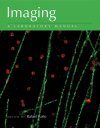
In recent years, imaging has rapidly become a tremendously valuable approach in nearly every field of biological research. Finding the right method and optimizing it for data collection can be a daunting process, even for an established imaging laboratory. Imaging: A Laboratory Manual is the cornerstone of a new laboratory manual series, designed as an essential guide for investigators who need these visualization techniques. This first volume is meant as a general reference for all fields, and describes the theory and practice of a wide array of imaging methods. From the basic chapters on optics, equipment and labeling to detailed explanations of advanced, cutting-edge methods like PALM, STORM, light sheet and high speed microscopy, Imaging: A Laboratory Manual is a vital resource for the modern biology laboratory.
Contents
Preface to the Book Series
Preface to Book 1
SECTION 1: INSTRUMENTATION
1. Microscope Principles and Optical Systems
Frederick Lanni and H. Ernst Keller
2. Video Microscopy, Video Cameras, and Image Enhancement
Masafumi Oshiro, Lowell A. Moomaw, and H. Ernst Keller
3. Differential Interference Contrast Imaging of Living Cells
Noam E. Ziv and Jackie Schiller
4. Infrared Video Microscopy
Hans-Ulrich Dodt, Klaus Becker, and Walter Zieglgänsberger
5. Confocal Microscopy: Principles and Practice
Alan Fine
6. Spinning-Disk Systems
Tony Wilson
7. Introduction to Multiphoton-Excitation Fluorescence Microscopy
Winfried Denk
8. How to Build a Two-Photon Microscope with a Confocal Scan Head
Volodymyr Nikolenko and Rafael Yuste
9. Arc Lamps and Monochromators for Fluorescence Microscopy
Rainer Uhl
10. Light-Emitting Diodes for Biological Microscopy
Tomokazu Sato and Venkatesh N. Murthy
11. Lasers for Nonlinear Microscopy
Frank Wise
12. Spectral Methods for Functional Brain Imaging
David Kleinfeld and Partha P. Mitra
13. Preparation of Cells and Tissues for Fluorescence Microscopy
Andrew H. Fischer, Kenneth A. Jacobson, Jack Rose, Pascal Lorentz, and Rolf Zeller
SECTION 2: LABELING AND INDICATORS
14. Labeling Cell Structures with Nonimmunological Fluorescent Dyes
Brad Chazotte
15. Introduction to Immunofluorescence Microscopy
George McNamara, Leonid (Alex) Belayev, Carl Boswell, and Jose Santos Da Silva Figueira
16. Immunoimaging: Studying Immune System Dynamics Using Two-Photon Microscopy
Melanie P. Matheu, Michael D. Cahalan, and Ian Parker
17. Biarsenical Labeling of Tetracysteine-Tagged Proteins
Asit K. Pattnaik and Debasis Panda
18. Bimolecular Fluorescence Complementation (BiFC) Analysis of Protein Interactions and Modifications In Living Cells
Tom K. Kerppola
19. Preparation and Use of Retroviral Vectors for Labeling, Imaging, and Genetically Manipulating Cells
Ayumu Tashiro, Chunmei Zhao, Hoonkyo Suh, and Fred H. Gage
20. Nonviral Gene Delivery
David A. Dean and Joshua Z. Gasiorowski
21. Cellular Bioluminescence Imaging
David K. Welsh and Takako Noguchi
22. Introduction to Indicators Based on Fluorescence Resonance Energy Transfer
Roger Y. Tsien
23. How Calcium Indicators Work
Stephen R. Adams
24. Calibration of Fluorescent Calcium Indicators
Fritjof Helmchen
25. Quantitative Aspects of Calcium Fluorimetry
Erwin Neher
26. Genetic Calcium Indicators: Fast Measurements Using Yellow Cameleons
Atsushi Miyawaki, Takeharu Nagai, and Hideaki Mizuno
27. Targeted Recombinant Aequorins
Tullio Pozzan and Rosario Rizzuto
28. Imaging Intracellular Signaling Using Two-Photon Fluorescent Lifetime Imaging Microscopy
Ryohei Yasuda
29. Imaging Gene Expression in Live Cells and Tissues
Hao Hong, Yunan Yang, and Weibo Cai
30. Multiphoton Excitation of Fluorescent Probes
Chris Xu and Warren R. Zipfel
SECTION 3: ADVANCED MICROSCOPY
Molecular Imaging
31. Single-Molecule FRET Using Total Internal Reflection Microscopy
Jeehae Park and Taekjip Ha
32. Alternating Laser Excitation for Solution-Based Single-Molecule FRET
Achillefs Kapanidis, Devdoot Majumdar, Mike Heilemann, Eyal Nir, and Shimon Weiss
33. FIONA: Nanometer Fluorescence Imaging
Paul D. Simonson and Paul R. Selvin
34. Photoactivated Localization Microscopy (PALM): An Optical Technique for Achieving~10-nm Resolution
Haining Zhong
35. Stochastic Optical Reconstruction Microscopy: A Method for Superresolution Fluorescence Imaging
Mark Bates, Sara A. Jones, and Xiaowei Zhuang
36. Imaging Live Cells Using Quantum Dots
Jyoti K. Jaiswal and Sanford M. Simon
37. Imaging Biological Samples with Atomic Force Microscopy
Pedro J. de Pablo and Mariano Carrión-Vázquez
Cellular Imaging
38. Total Internal Reflection Fluorescence Microscopy
Ahmet Yildiz and Ronald D. Vale
39. Fluorescence Correlation Spectroscopy: Principles and Applications
Kirsten Bacia, Elke Haustein, and Petra Schwille
40. Image Correlation Spectroscopy: Principles and Applications
Paul W. Wiseman
41. Time-Domain Fluorescence Lifetime Imaging Microscopy: A Quantitative Method to Follow Transient Protein–Protein Interactions in Living Cells
Sergi Padilla-Parra, Nicolas Audugé, Marc Tramier, and Maïté Coppey-Moisan
42. Single- and Two-Photon Fluorescence Recovery after Photobleaching
Kelley D. Sullivan, Ania K. Majewska, and Edward B. Brown
43. Fluorescent Speckle Microscopy
Lisa A. Cameron, Benjamin R. Houghtaling, and Ge Yang
44. Polarized Light Microscopy: Principles and Practice
Rudolf Oldenbourg
45. Array Tomography: High-Resolution Three-Dimensional Immunofluorescence
Kristina D. Micheva, Nancy O’Rourke, Brad Busse, and Stephen J Smith
46. Monitoring Membrane Potential with Second-Harmonic Generation
Stacy A. Wilson, Andrew Millard, Aaron Lewis, and Leslie M. Loew
47. Grating Imager Systems for Fluorescence Optical-Sectioning Microscopy
Frederick Lanni
Tissue Imaging
48. Coherent Raman Tissue Imaging in the Brain
Brian G. Saar, Christian W. Freudiger, Xiaoyin Xu, Anita Huttner, Santosh Kesari, Geoffrey Young, and X. Sunney Xie
49. Ultramicroscopy: Light-Sheet-Based Microscopy for Imaging Centimeter-Sized Objects with Micrometer Resolution
Klaus Becker, Nina Jährling, Saiedeh Saghafi, and Hans-Ulrich Dodt
50. In Vivo Optical Microendoscopy for Imaging Cells Lying Deep within Live Tissue
Robert P.J. Barretto and Mark J. Schnitzer
51. Light-Sheet-Based Fluorescence Microscopy for Three-Dimensional Imaging of Biological Samples
Jim Swoger, Francesco Pampaloni, and Ernst H.K. Stelzer
52. Photoacoustic Imaging
Yin Zhang, Hao Hong, and Weibo Cai
Fast Imaging
53. Imaging the Dynamics of Biological Processes via Fast Confocal Microscopy and Image Processing
Michael Liebling
54. High-Speed Two-Photon Imaging
Gaddum Duemani Reddy and Peter Saggau
55. Digital Micromirror Devices: Principles and Applications in Imaging
Vivek Bansal and Peter Saggau
56. Spatial Light Modulator Microscopy
Volodymyr Nikolenko, Darcy S. Peterka, Roberto Araya, Alan Woodruff, and Rafael Yuste
57. Temporal Focusing Microscopy
Dan Oron and Yaron Silberberg
Uncaging
58. Caged Neurotransmitters and Other Caged Compounds: Design and Application
George P. Hess, Ryan W. Lewis, and Yongli Chen
59. Nitrobenzyl-Based Caged Neurotransmitters
Graham C.R. Ellis-Davies
60. Uncaging with Visible Light: Inorganic Caged Compounds
Leonardo Zayat, Luis M. Baraldo, and Roberto Etchenique
SECTION 4: APPENDICES
1. Electromagnetic Spectrum
Marilu Hoeppner
2. Fluorescence Microscopy Filters and Excitation/Emission Spectra
3. Safe Operation of a Fluorescence Microscope
George McNamara
4. Microscope Objective Lenses
5. Glossary
6. Cautions
Index
Customer Reviews


![Manuale di Microscopia dei Funghi [Manual to Microscopy of Fungi]](http://mediacdn.nhbs.com/jackets/jackets_resizer_medium/26/260752.jpg?height=150&width=104)

![Manuale di Microscopia dei Funghi, Volume 2 [Manual to Microscopy of Fungi, Volume 2]](http://mediacdn.nhbs.com/jackets/jackets_resizer_medium/23/236960.jpg?height=150&width=107)


